One of the first steps toward good eye care and healthy eyes is an eye exam. Did you know that a routine eye exam involves more than correcting your vision with glasses or contacts? Read on about how eye exams assess vision health and the different types of tests.
What Is an Eye Exam or Vision Test?
An eye exam is a group of vision tests that an eye doctor (optometrist or ophthalmologist) uses to examine the health of your eyes. Routine eye exams are important and can detect serious vision problems like glaucoma, cataracts and macular degeneration so they can be treated at an early stage. In fact, a comprehensive eye exam is less about your eyeglass or contact lens prescription and more about your overall eye health.
Vision tests are the basic parts of an eye examination, but during a typical eye exam, you'll also discuss any recent vision changes you may have noticed. Don't hesitate to ask questions if you don't understand something during your appointment. Then, make sure to keep scheduling routine eye exams to maintain healthy vision.
8 Routine Eye Exam Tests
Below are eight types of eye tests that are usually part of a routine eye exam, complete with an in-depth description that includes national average costs* and cost ranges.
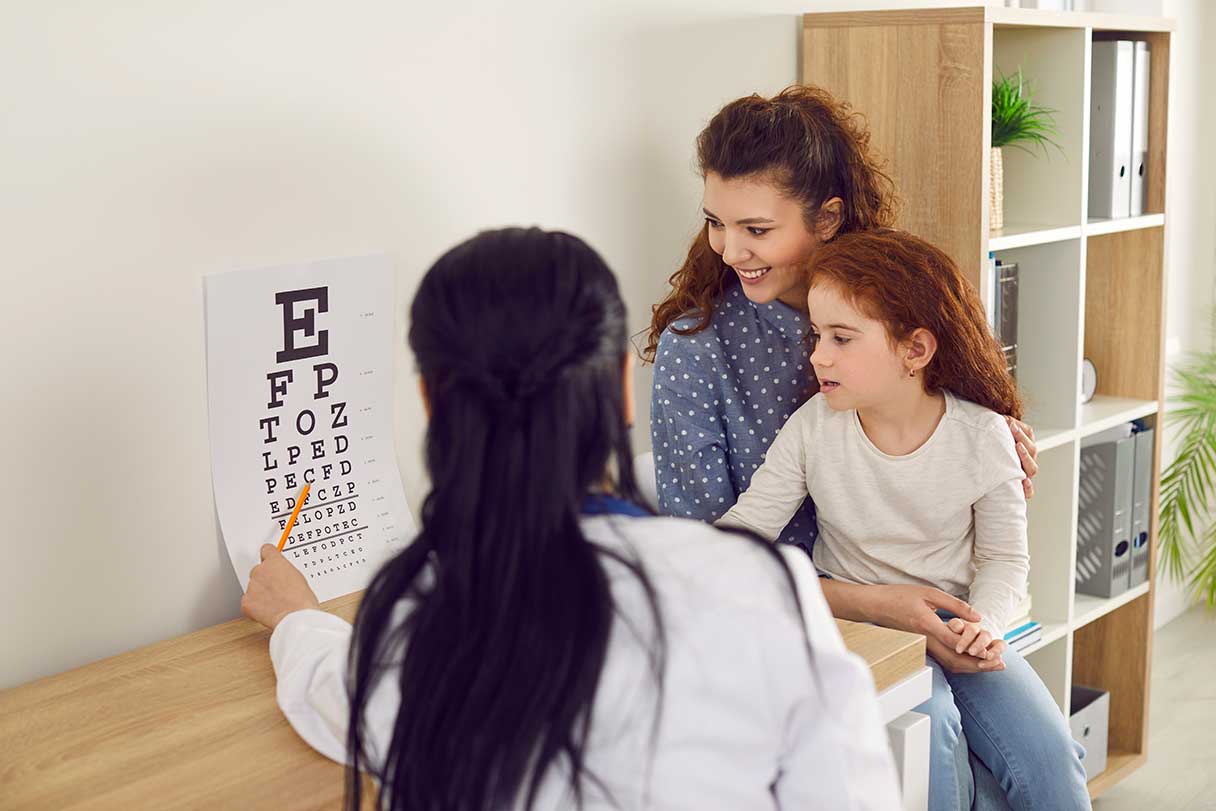
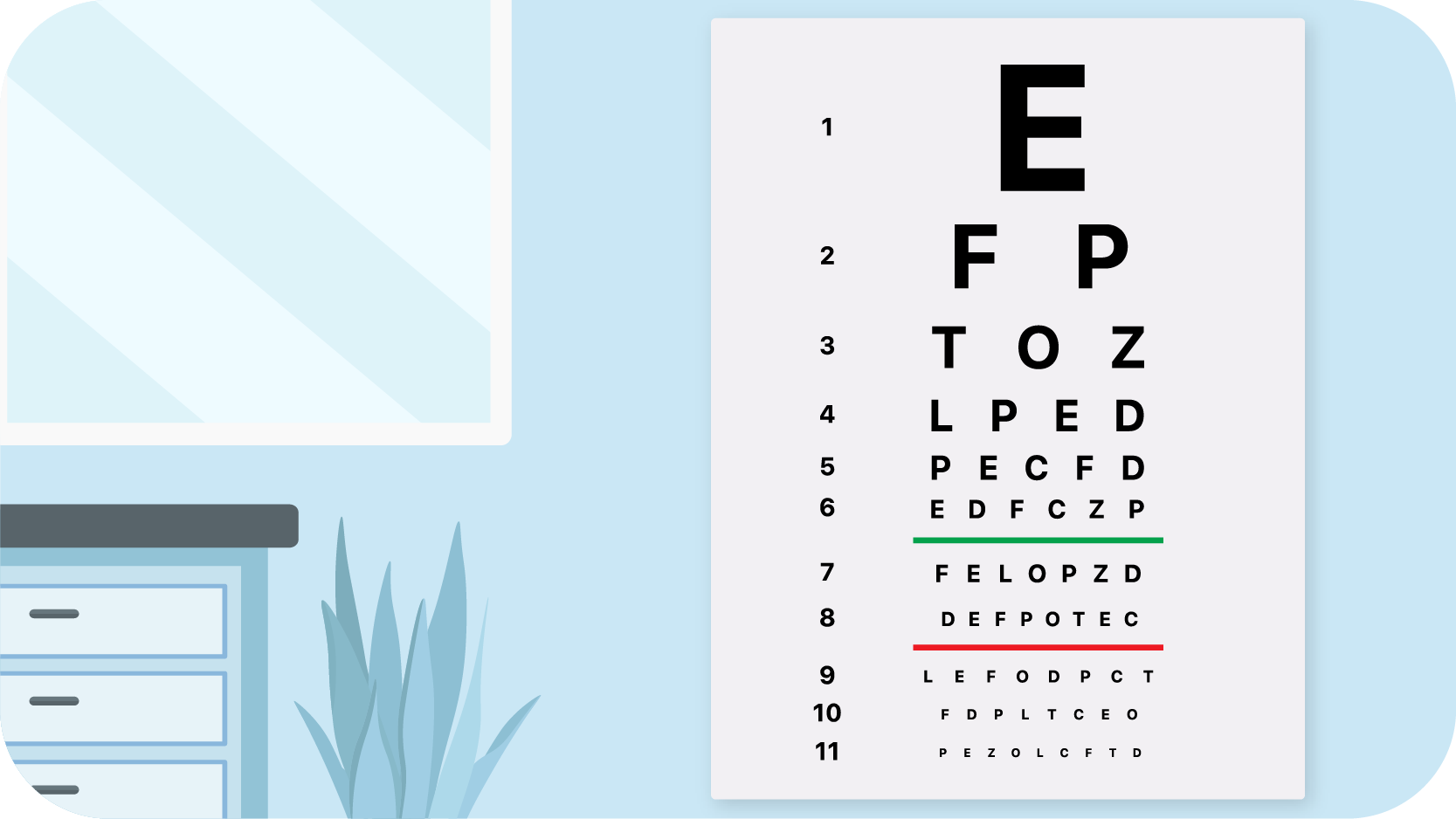
1. Visual Acuity Test
This is the "eye chart" test that you may be familiar with. A visual acuity test measures the ability of each individual eye to distinguish shapes, such as letters or numbers, at a given distance.
You're positioned about 20 feet away from the chart and asked to identify a series of letters or numbers printed on a chart (officially called a Snellen Eye Chart). The lines of type get smaller as you move down the chart. Each eye is tested individually, and it's an important test that done on a consistent basis can track changes in vision.
Cost of visual acuity test
The average cost of a visual acuity test is $25, with a range between $20 and $49.1
2. Visual Refraction Eye Test
Refraction refers to the way light waves are bent as they pass through your cornea and lens. A refraction assessment helps your eye doctor determine if you need vision correction, as well as the corrective lens prescription that will help give you the sharpest, clearest vision.
Your vision provider may use a device called a phoropter to help determine what strength of prescription lenses will correct your refractive error. This exam is performed in a dark room to help you better view the images through the phoropter lenses. Repeating this step several times helps your provider find the lenses that can give you the best possible vision.
Cost of visual refraction eye test
A visual refraction eye test costs $46 on average but can range from $36 to $87.1
3. Visual Field Test
This test helps determine your central field of vision, which is the area right in front of you, or what you can see without moving your eyes. It also checks your peripheral vision, which is the area above, below and to the sides of your central vision.
There are several types of visual field tests used to assess different aspects of eye function:
- Binocular vision test. Evaluates how well the eyes work together
- Developmental vision evaluation. Assesses how vision problems may affect learning
- Frequency doubling technology (FDT). Detects potential blind spots
- Goldmann kinetic perimetry. Measures visual field using moving stimuli
- Standard automated perimeter (SAP). The most common test, where a computer flashes small lights inside a bowl-shaped device; you press a button each time you see a light to measure your field of vision
Cost of visual field test
The average cost of a standard automated perimetry visual field test is $109 but can range from $84 to $186.1
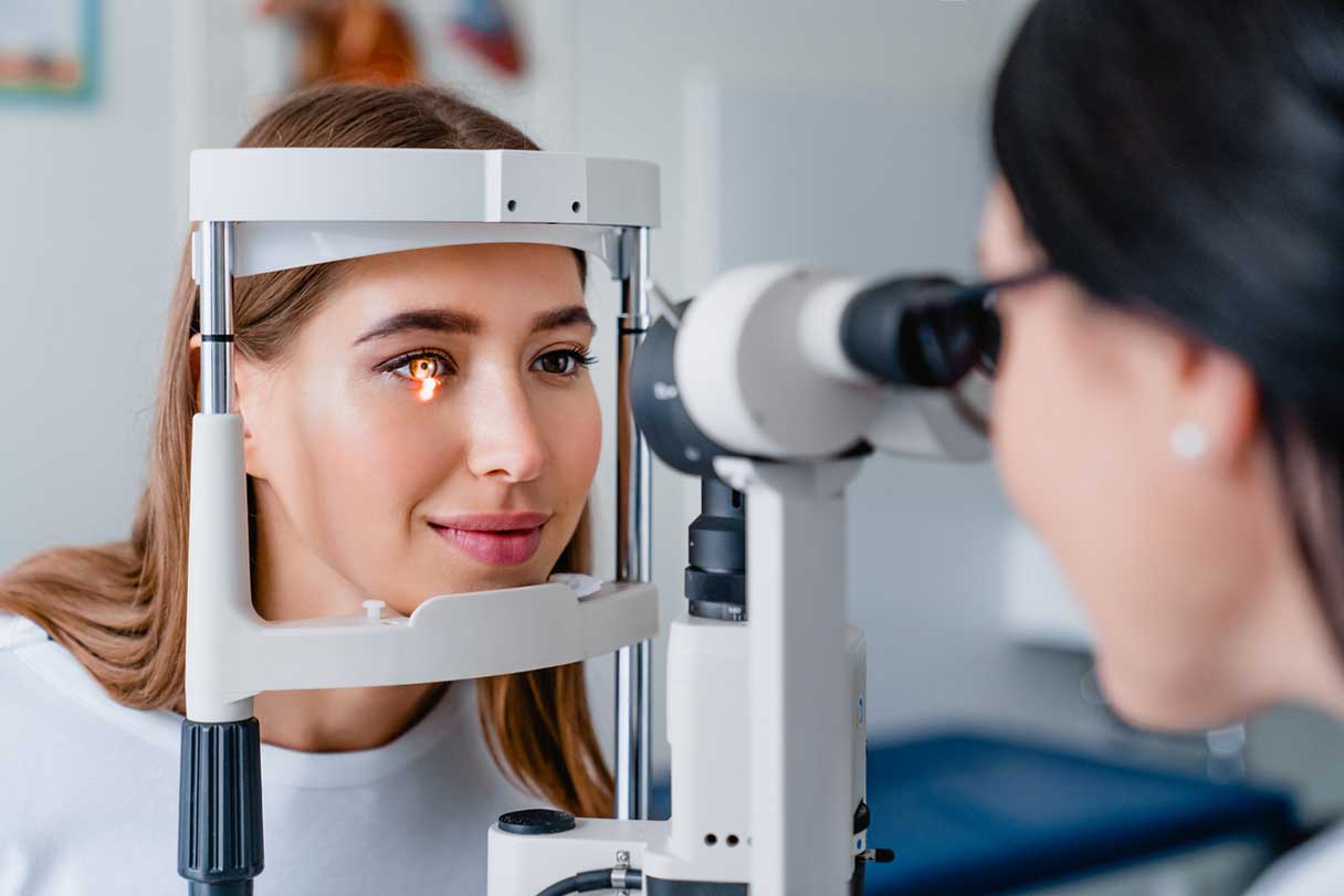
4. Slit-Lamp Exam
A slit lamp is a microscope that lets your doctor use intense light to enhance the view of the front of your eye, so your doctor can examine the cornea, iris, lens and anterior chamber. When examining your cornea, your provider may use eye drops containing fluorescein dye. This orange dye makes it easier for your eye doctor to look for areas of dryness, small cuts, scrapes, tears, foreign objects or infections on your cornea. As you blink, your natural tears will rinse the dye away.
Cost of slit-lamp test
A slit lamp test costs $55 on average but can range from $42 to $100.1
5. Glaucoma Test
This exam measures the pressure inside your eye, referred to as intraocular pressure. It helps your eye doctor detect glaucoma, a disease that causes pressure to build up inside your eyes and can cause blindness. Glaucoma can be treated if it's caught early.
The most common test for eye pressure today is a tonometry test, which is a device that applies a gentle puff of air onto the eye's surface to test the pressure. Other glaucoma tests include perimetry, which maps the peripheral vision to detect blind spots, and gonioscopy, which examines the angle where the iris meets the cornea to ensure it's open and free of blockages.1
Cost of glaucoma test
The average cost for a tonometry glaucoma test is $21, but can range between $16 and $40.1
6. Color Vision Test
This test screens for color blindness by examining your ability to distinguish colors. You might be asked to detect a number or letter within an image of multicolored circles. This is an important test, since certain professions like law enforcement or pilots require perfect color vision.
Cost of color vision test
A color blindness test costs $50 on average but can range from $38 to $96.1
7. Corneal Topography
These test for variations of the curvature of the surface of your corneas. While you stare at an object, your eye doctor takes thousands of pictures of the surface of your eye to map the shape of your cornea. This can help them see if your cornea has an irregular curvature, which is called astigmatism. It also helps your doctor create contact lenses that fit your eye, and can help them prepare for things like cataract surgery, LASIK or corneal transplant surgery.
The most common and standard method involves using a Placido disc to project concentric rings onto the cornea. The reflected rings are then captured and analyzed to create a detailed map of the cornea's surface.1
Cost of corneal topography
The average cost of a Placido disc corneal topography test is $182 but can range from $140 to $299.1
8. Ophthalmoscopy (Fundoscopy)
Your doctor will put special eye drops in your eyes to dilate them (increase the size of your pupil). You'll be asked to wait 15 to 20 minutes for the solution to take effect. You may notice during this time that light seems much brighter. Then, your doctor will shine a light into your eye. This test helps them get a better look at your optic nerve, lens and retina.
Direct ophthalmoscopy uses a handheld device to provide a highly magnified but narrow view of the back of the eye. Indirect ophthalmoscopy, with a head-mounted binocular microscope and handheld lens, offers a wider view, making it ideal for examining peripheral areas.1
Cost of an ophthalmoscopy (fundoscopy)
The average cost of an indirect ophthalmoscopy is $130, with a range of $100 to $224. A direct ophthalmoscopy costs $104 on average but can range from $80 to $200.1
Why Eye Exams Are Recommended
Many adults, especially those in their 30s and 40s, forgo vision tests because they think they don't need them. But eye exams can help you get out in front of blindness-causing conditions like glaucoma, where early diagnosis and treatment can slow vision loss.
Eye tests can help keep your eyes healthy by assessing:
- How clearly you see
- If one eye has better vision than the other
- If you have astigmatism or color blindness
- Your peripheral vision
- Whether you have a condition that needs treatment (such as glaucoma, cataracts or macular degeneration)
In addition to strictly vision-related conditions, eye tests may also help doctors identify other conditions like:
- Aneurysms
- Brain tumors
- Cancer
- Diabetes
- Heart disease
- High blood pressure
- High cholesterol
- Lupus
- Lyme disease
- Multiple sclerosis
- Sexually transmitted diseases
- Stroke
Financing Eye Exams With the CareCredit Credit Card
Regular eye exams are an important part of your eye health. Whether you're getting an eye exam or paying for eye surgery, the CareCredit credit card can help you pay for care where your insurance leaves off.** Use our Acceptance Locator to find a vision specialist near you that accepts CareCredit. Continue your wellness journey by downloading the CareCredit Mobile App to manage your account, find a provider on the go and easily access the Well U blog for more great articles, podcasts and videos.
In addition to vision care, you can also use your CareCredit credit card for dentistry, cosmetic, pet care, hearing, health systems, dermatology, pharmacy purchases, spa treatments and so much more within the CareCredit network. How will you invest in your health and wellness next?
Expert Reviewer
Dr. Alexandra Chebil, M.D., F.R.C.S.C.
Dr. Alexandra Chebil is a board-certified ophthalmologist with the Lasik Center Medical Group in Newport Beach, California, who has more than 25 years of experience with refractive surgery and noninvasive facial aesthetic procedures. She was one of the first ophthalmologists to perform LASIK, and has successfully performed more than 70,000 procedures.
Author Bio
Stephanie Dwilson specializes in science journalism, breaking news and animal health and is a business owner, non-practicing attorney and writer.
Related Tags
Want to share this on social media?